
Method for recording bioelectrical signals in fungal mycelia
New sonification devices claim to make the well-being and communication of certain living organisms audible. This article examines how a state-of-the-art biodata sonification circuit works, what signals it can detect, and how it converts those signals.
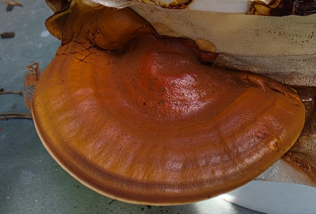
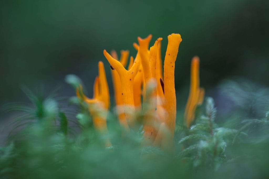
Furthermore, an alternative, more scientific approach to recording, visualising, and converting bioelectric signals from the mycelium of the Ganoderma lucidum fungus (Figure 1) is explored and illustrated, and its possibilities discussed.
In recent years, devices advertised as capable of converting the bioelectrical signals of plants and fungi into live sonic experiences have been developed. These devices, which are primarily used by artists and musicians, are supposedly able to make the well-being of, and even the communication between, living organisms audible in real time.
The idea of experimenting with such a device arose during research into possible applications of Ganoderma lucidum mycelium as a building material for small structuresat South-Eastern Finland University of Applied Sciences – Xamk.
The sonification device built and used for the initial experiments is based on an astable multivibrator circuit that converts the changing currents resulting from changes in the studied specimen’s resistance into a square wave. The frequency of the wave at the circuit’s output changes with the input current.
The period of that signal is subsequently detected by a microprocessor, which generates control signals for a sound source using mathematical operations such as averaging and standard deviation (Cusumano n.d.). In other words, the device does not actually hear the organism. Instead, it turns tiny electrical changes in a living organism into signals that a computer can read and use.
In the initial tests, the device produced an interesting sonic experience when the electrodes were embedded in mycelium, attached to plant leaves or the human body. However, when the electrodes were attached to a wet paper towel, the sounds generated were equally intriguing (Figure 2). Further laboratory tests showed that even when a fixed resistor was attached to the electrodes, the device still produced similar melodies, suggesting that the circuit is highly sensitive and that the sounds generated are mostly due to electromagnetic noise rather than the bioelectrical signals of the test specimens.
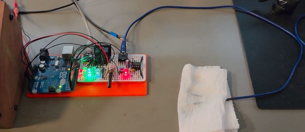
Having set out to use the mycelium’s actual bioelectric signals to create sound (Bircher 2026), the findings from a wet paper towel indicated that the current method was interesting but ultimately erroneous. As previous research has shown that electrical signals are present in growing mycelium and can be recorded (Buffi et al. 2025), it was necessary to adopt a more scientific approach to studying the electrical activity in growing mycelium.
Recording electrical activity in growing mycelium
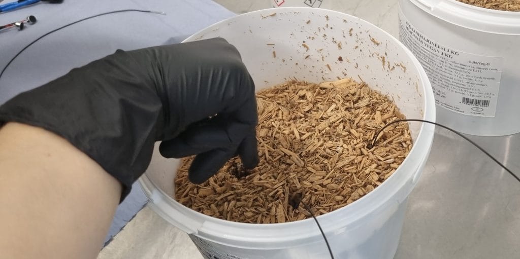
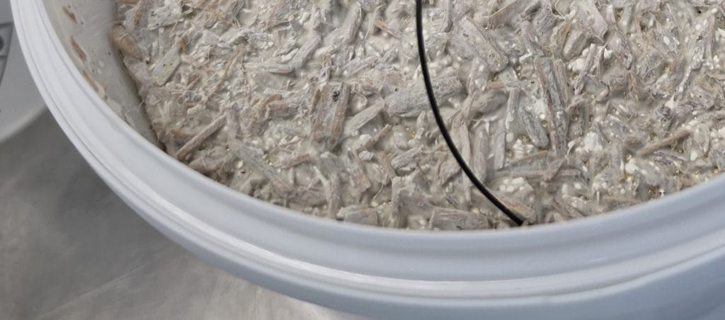
A 6 ½-digit lab-grade test instrument was connected to wires with striped ends, which were placed in a plastic bucket filled with straw based substrate and served as electrodes (Figure 3). To ensure that the electrodes made contact with the mycelium, the resistance measurement was initiated as soon as the mycelium became visible on the substrate’s surface (Figure 4).
The test instrument was configured to log one data point every four seconds, with an integration time of 100 power cycles (lasting two seconds) to filter out higher-frequency noise, especially from 50 Hz power lines. The logged data points had to be downloaded periodically before the ring buffer overflowed.
The files containing the results were merged and formatted using regular expressions.
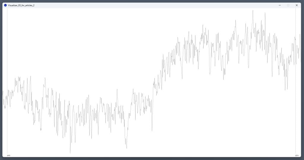
A custom application was written in Java to display the data points as a curve and to magnify regions of interest (Figure 5).
Review of the Data
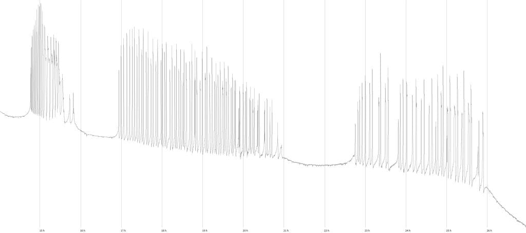
Over 57 hours, 51487 consecutive data points were collected (Figure 6). During the first 14 hours of the recorded data, only one outlier (Region 1 in Figure 6) occurred: a spike with elevated resistance lasting about 6 minutes. Thereafter, 11 hours of strong activity were detected (Region 2 in Figure 6; Figure 7), during which three bursts with 16 to 18 spikes per hour appeared. A further cluster of activity (Region 3 in Figure 6) is apparent after 5,5 hours of relative inactivity. The recording was terminated after 23 hours of inactivity.
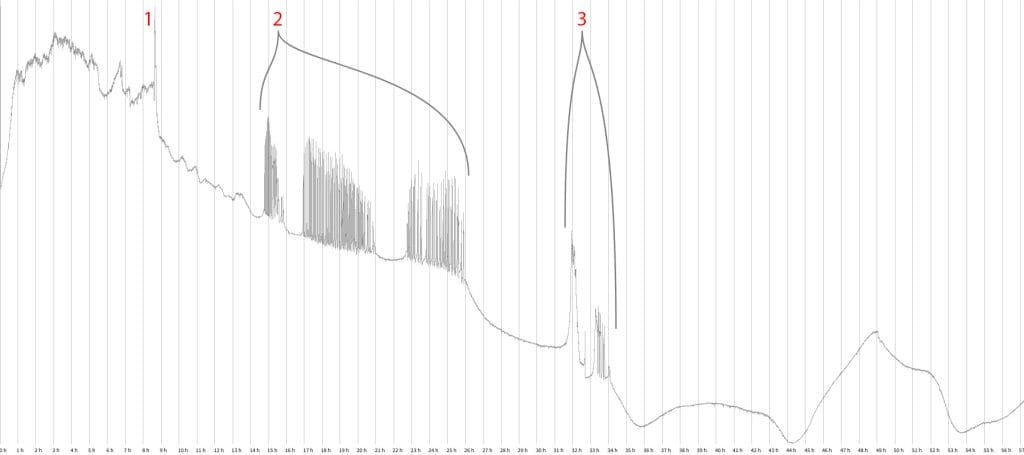
Similar spikes have been detected earlier in oyster fungi by Adamatzky (2018); however, they measured voltage in their experiments. In our case, the general downward trend in resistance (Figure 6) may be attributed to increased mycelial content in the substrate, which in turn increases the conductivity of the material between the electrodes.
It has to be noted that the measured resistance is well within the bandwidth of the sonification device initially tested. Because the recorded signals are relatively slow and there are prolonged periods of low activity, the sonification device would, however, not be able to create an interesting sonic experience based on signals that clearly originate from the species under test (Bircher 2026).
Lessons learned
These experiments were successful, as we set out to detect and record bioelectric signals that can serve as components in creating music. The strength of the low-frequency components of the recorded signals and their similarity to the spiking behaviour detected in other research suggest that they originate from the mycelium itself. However, in these exploratory experiments, no attention was paid to logging other data for comparison with our results.
The test instrument required manual downloading of the data every five hours, which required planning and dedication. For future research, a lab-grade multichannel data acquisition solution would be preferable, allowing for recording at higher frequencies and over longer periods.
While the makeshift software application used in these experiments was adequate, there are certainly better tools for viewing and potentially analysing the results, and for combining different datasets to examine correlations.
These experiments were conducted using a mix of creative and technological skills and mindsets. It will be interesting to see what other disciplines, such as biology and data science, can contribute to similar research in the future.
Outlook
What started as a creative endeavour led to the realisation that it is possible to record electrical signals originating from the mycelium of Ganoderma lucidum, and the resulting waveforms are clearly more than just random noise.
There is considerable potential for future research in which not only the electrical signals but also the mycelial growth, health, and environmental factors are logged. This might yield useful information about optimal growing conditions and the mycelium’s response to unfavourable or even harmful conditions, such as mould infestations.
Currently an increased research interest into mycelium as a sustainable component for e.g. the construction and packaging industry can be observed. Continuing our research could support such endeavours and help to further understand this interesting material.
These experiments were conducted as part of the “Experimental environments for climate-responsible outdoor structures” project, in which the use of mycelium plays a pivotal role. The project is funded by the Regional Council of Kymenlaakso and co-funded by the European Union.
The Author has examined the research featured in this article in a second publication, which highlights the creative possibilities of the experiments conducted (Bircher 2026).
Read also
Bircher, M. 2026. Can mushrooms sing? Bioelectrical signals as a component in creative work.
Lähteet
Adamatzky, A. 2018. On Spiking Behaviour of Oyster Fungi Pleurotus Djamor. Scientific Reports 8, 7873. Available at: https://doi.org/10.1038/s41598-018-26007-1.
Bircher, M. 2026. Can mushrooms sing? Bioelectrical signals as a component in creative work. Elo publication channel. Southeastern Finland University of Applied Sciences. Available at: https://urn.fi/URN:NBN:fi-fe20261001130311
Buffi, M., Kelliher, J., Robinson, A., Gonzalez, D., Cailleau, G., Macalindong, J., Frau, E., Schintke, S., Chain, P., Stanley, C., Künzler, M., Bindschedler, S. & Junier, P. 2025. Electrical Signaling in Fungi: Past and Present Challenges. FEMS Microbiology Reviews 49. Available at: https://doi.org/10.1093/femsre/fuaf009.
Cusumano, S. No date. Biodata Sonification. Electricity for Progress. Available at: https://electricityforprogress.com/ [Accessed: 14.6.2026].
Lisenssi
Viittausohjeet
Bircher, M. 2026. Method for recording bioelectrical signals in fungal mycelia. Kesto online publication. Available at: https://urn.fi/URN:NBN:fi-fe20260923128292.